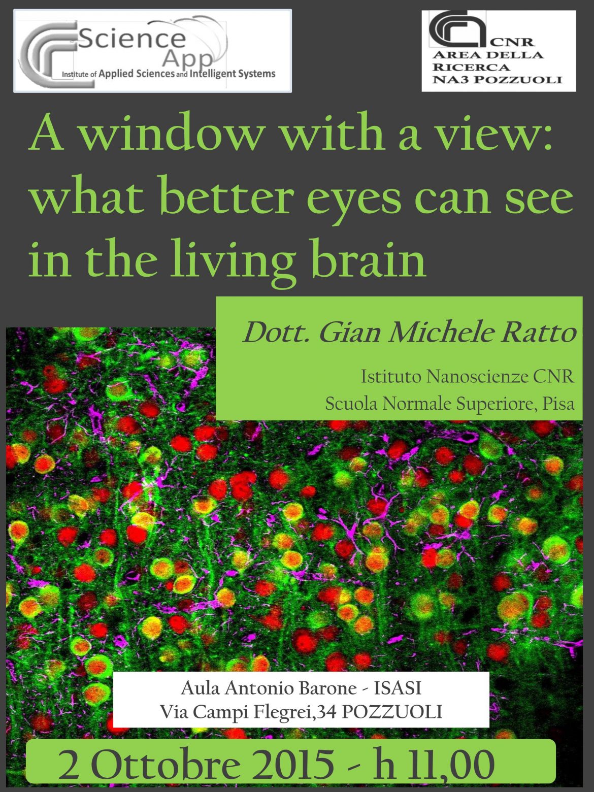
Two photon micr oscopy has emerged as a powerful tool to image form and function in the brain in vivo: neurons and glia can be visualised by means of an ever expanding range of fluorescent probes and a wide variety of information can be collected in the intact brain. For example, transgenic mice expressing the Green Fluorescent Protein in selected neuronal population can be used to image the morphological development of neurons. Functional studies relies on genetically encoded or synthetic dyes to visualize cellular and network function with sub cellular resolution. At this time, two photon imaging is the only available tool that allows to peer into neuronal function in the intact brain at the cell population level. Indeed, intravital imaging has provided novel insights on brain structure and function with unprecedented temporal and spatial resolution. The availability of genetic and pharmacological models of brain pathologies, allow to investigate function and form in the normal and diseased cortex. In this talk I will give an overview of what we can see in the brain when we put together two photon imaging with novel genetic sensor to image different aspects of brain cells.
First, I will show a molecular tool that allow to visualize in great details neuronal connectivity in a mosaic model of brain disease. In brief, I will show the operation of a fluorescent sensor/effector that can be used to create a mosaic of neurons expressing two alternative genetic backgrounds that are reported to the external world by two different fluorescent proteins. I will discuss briefly the implications of this tool for the study of brain disease and in oncology.
Next, I will show how a genetically encoded sensor can be used to measure pH and Chloride concentration in vivo. The 2-photon spectral properties of the sensor, show that changes in chloride and pH cause changes in the emission and excitation spectra of the sensor. Then, we found that brain tissue has a strongly wavelength-dependent effect on the propagation of excitation and emission strongly affecting the pH and Chloride measures. Finally, we demonstrate that by means of analytical spectral decomposition we can measure and compensate for the spectral distortion caused by the tissue. Our data are the first direct measure of intracellular chloride ever attained in vivo.

