
Description of the activity
The possibility to obtain three-dimensional quantitative information of biological samples in a label-free and non-invasively way is of fundamental importance to understand the inner workings of cells and their healthy state. Moreover, optical properties of the sample, such as refractive index distribution and state of polarization, could give additional information, as many biological materials are birefringent.
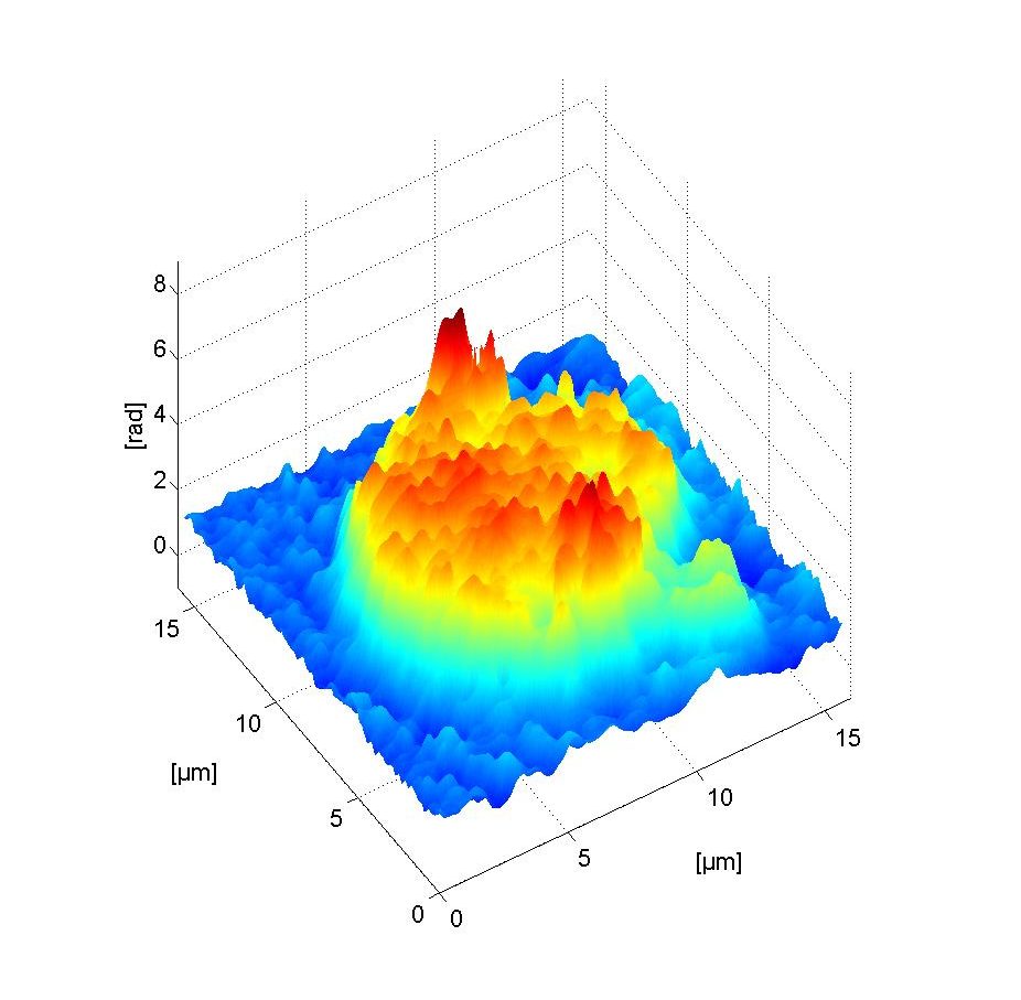
Polarization sensitive digital holographic imaging (PSDHI) is an emerging technique permitting to achieve not only quantitative morphological information, such as the classic digital holography imaging, but also intrinsic information about the polarization state of the sample through the phase change quantification.
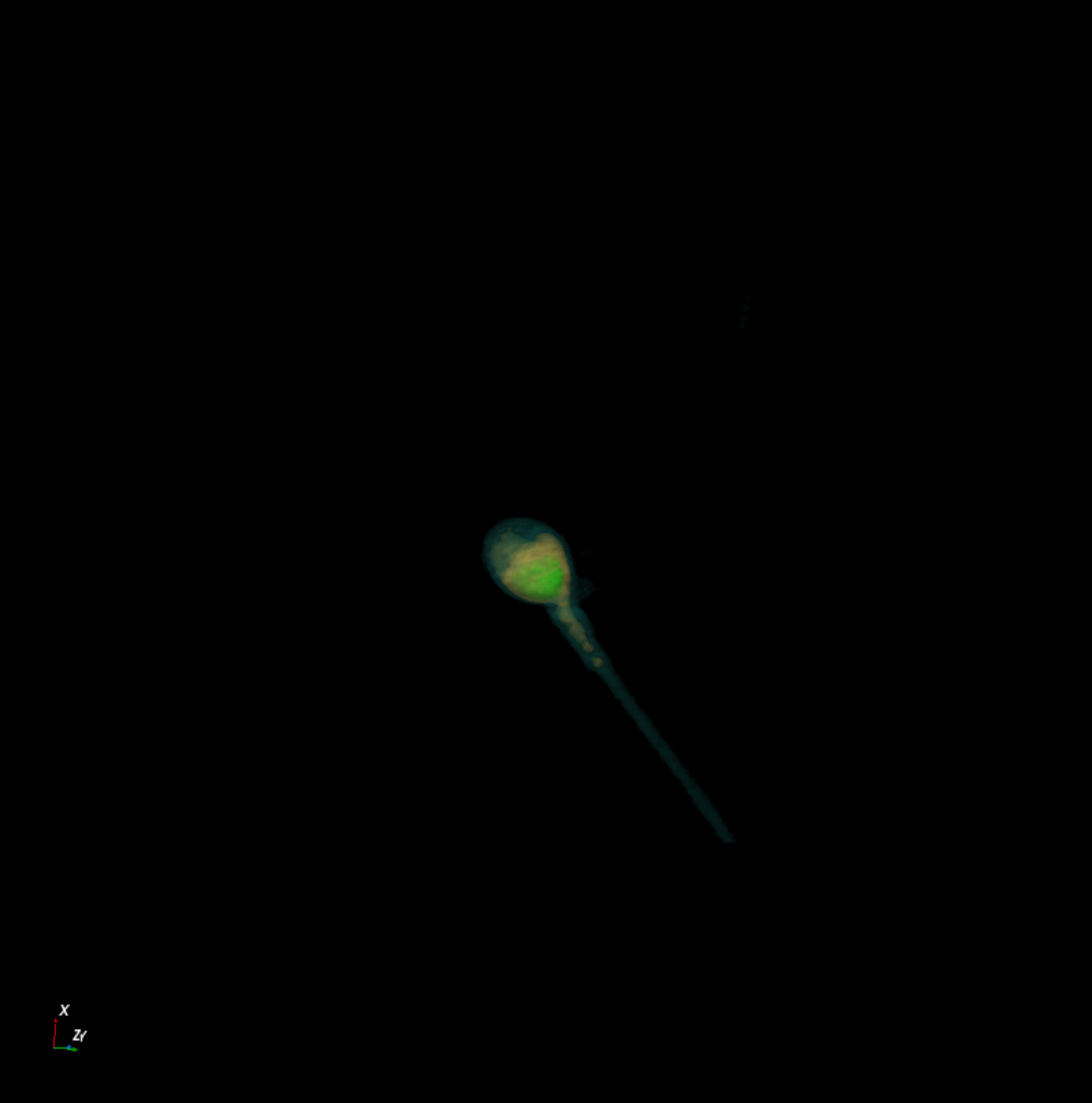
The research activity aims to take advantage of PSDHI technique to classify the state of maturation of healthy human spermatozoa and to differentiate cancer cells from healthy cells belonging to the same tissue or organ.
Involved personnel
National and International Collaborations
- CNR (Institute Experimental Endocrinology and Oncology – IEOS)
- Centro di Fecondazione Assistita Napoli (IVF)
Instrumentation/facilities
- Homemade polarization sensitive digital holographic imaging setup which includes: an inverted microscope; high coherent visible laser sources; polarized-sensitive optical fibers; microscope objective lenses for quantitative polarization imaging; CCD.
- Tomocube HT-2H: Holotomographic microscopy with 3D fluorescence imaging capability equipped with temperature controllers and automatic gas mixer for in vivo acquisitions.