Description of the activity
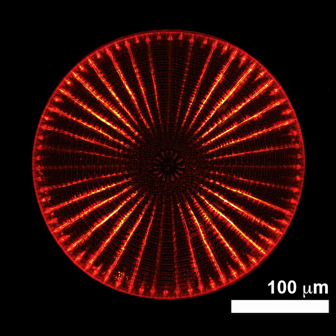
Several organisms including algae, flora, insects, and birds, are provided with sub-micrometric structures able to manipulate light with high efficiency, mainly for intra- and inter-species communication. Some of these periodic or quasi-periodic structures can cause coherent or incoherent scattering; others act as one-dimensional, spectrally selective multilayer reflectors, polarization-selective reflectors, two-dimensional diffraction gratings, and photonic crystals, and can be shaped in complex hierarchical architectures whose optical properties rely on a fine interplay between order and disorder. In particular, we study the photonic properties of diatoms, unicellular microalgae which represent the main component of phytoplankton. Their protoplasm is enclosed in a regularly micro- and nano-patterned silica shell, the frustule, which presents some analogies with artificial photonic crystals. We observed several properties (e.g. diffractive-induced light confinement, photoluminescence, manipulation of polarization, UV-R screening) and exploited them in the realization of bio-based super-lensing, bio-derived SERS substrates, optical biosensors, and dielectric metasurfaces, to name a few. These studies are conducted by means of high-performance numerical simulations, transmission and fluorescence imaging, digital holography, photoluminescence spectroscopy, and interaction with structured light. Optical characterization has been recently extended also to living organisms.

Involved personnel
E. De Tommasi | M.A. Ferrara | L. De Stefano | I. Rea
National and International Collaborations
- Institute of Endocrinology and Experimental Oncology (IEOS), CNR;
- Institute of Biosciences and Bioresources (IBBR), CNR;
- Stazione Zoologica Anton Dohrn;
- University of Campania “Luigi Vanvitelli”;
- University of St Andrews, Scotland, UK;
- University of Gothenburg, Sweden;
- University of Antwerp, Belgium.
Instrumentation/facilities
- DELL workstation (Xeon bi-processor with 2-14 cores, 512 GB RAM, graphic board NVIDIA QUADRO K6000) for numerical simulations.
- Field Emission Scanning Electron Microscope (Carl Zeiss NTS GmbH 1500 Raith) and Atomic Force Microscope (XE-70 Park’s) for morphological characterization of diatom frustules.
- Laser sources at 406 nm (diode laser, Micro Laser Systems, L4-408-48B-TE), 532 nm (diode laser, Laserslt, DPSS), and 633 nm (He-Ne laser, Research Electro-Optics, HRP350-EC) for transmission characterization of diatom valves.
- UV-VIS fiber lamp (Hamamatsu, L10290) for optical characterization of diatom frustules with non-coherent light.
- UV optics and sensors for the study of the interaction of diatom frustules with UV-R.
- He-Cd laser source (Kimmon, IK5751I-G) plus spectrometer (Princeton Instruments, SpectraPro 300i) for photoluminescence characterization.
- Fluorescence microscope (Leica Microsystems, DM6M).
- Home-made digital holographic microscope (laser source at 660 nm).
- Liquid-crystal-on-silicon spatial light modulator (Hamamatsu, LCOS-SLM X13138) for the study of the interaction of diatom valves with structured light.
Active projects and contracts
- “Photonic properties of diatom frustules – functional meaning and beyond”, Swedish Research Council, grant number 2018-04289, in collaboration with University of Gothenburg (Sweden) and Università della Campania “Luigi Vanvitelli” (Italy). Principal investigator: Prof. Angela Wulff (University of Gothenburg, Sweden).